Cataract
What is Cataract?
A cataract is a clouding of the lens in the eye that affects vision. The lens must be clear for the retina to receive a sharp image. If the lens is cloudy from a cataract, the image you see will be blurred. The lens lies behind the pupil and works much a like a camera lens. It focuses light onto the retina at the back of the eye. Also, the lens adjusts the eye's focus, letting us see things clearly both up close and far away.
How can cataracts affect my vision?
The lens consists mostly of water and protein. Clumps of protein reduces the sharpness of the image. The clear lens also slowly changes to yellowish brown in color, adding a brownish tint to vision.
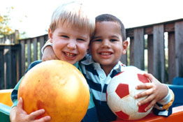
Normal vision:
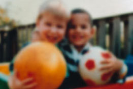
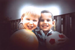
 Viewed by person with cataract:
Who is at risk for cataract?
People can get "age-related" cataracts in their 40s and 50s but they are mostly small and do not affect vision. It is after 60s that most cataracts severely affect the vision. Other risks includes Diabetes, smoking, and prolonged exposure to sunlight.
What can I do to protect my vision?
If you are 60 or older, you should have a comprehensive eye exam to detect cataracts. If you also suffer from diabetes, glaucoma, and had an eye injury you should be screened for cataracts. Wearing sunglasses and regular intake of antioxidants, such as green leafy vegetables, fruits, Vitamin A, C, E can help reduce the risk of age related cataracts.
How do you treat Cataract?
Cataract surgery is one of the most common operations performed in United States. Most surgery done is by phacoemulsification. The surgeon makes an incision on the side of cornea, and a tiny probe emits ultrasound waves that break up the lens so that it can be removed by suction. After the natural lens has been removed, it's replaced by an artificial lens (IOL). In most cases, healing will be complete within eight weeks. Problems after surgery are rare but infection, bleeding, blurry vision, or inflammation can occur. Sometimes the eye tissue surrounding the new artificial lens becomes cloudy and is treated with a laser. Your doctor will schedule eye exams to check your progress.
Glaucoma Defined
What is glaucoma?
Glaucoma is a group of diseases that can damage the eye's optic nerve and result in vision loss and blindness. Glaucoma occurs when the normal fluid pressure inside the eyes slowly rises. However, with early treatment, you can often protect your eyes against serious vision loss.
What are some other forms of glaucoma?
Open-angle glaucoma is the most common form. However, some people have other types of the disease which are:
- Low-tension or normal-tension glaucoma
- Angle-closure glaucoma
- Congenital glaucoma
- Secondary glaucoma
How does open-angle glaucoma damage the optic nerve?
In the front of the eye is a space called the anterior chamber. A clear fluid flows continuously in and out of the chamber and nourishes nearby tissues. The fluid leaves the chamber at the open angle where the cornea and iris meet. When the fluid reaches the angle, it flows through a spongy meshwork, like a drain, and leaves the eye. Sometimes, when the fluid reaches the angle, it passes too slowly through the meshwork drain. As the fluid builds up, the pressure inside the eye rises to a level that may damage the optic nerve. When the optic nerve is damaged from increased pressure, open-angle glaucoma--and vision loss--may result. That's why controlling pressure inside the eye is important.
Does increased eye pressure mean that I have glaucoma?
Not exactly. Increased eye pressure means you are at risk for glaucoma, but does not mean you have the disease. A person has glaucoma only if the optic nerve is damaged. If you have increased eye pressure but no damage to the optic nerve, you do not have glaucoma. However, you are at risk. Follow the advice of your eye care professional.
Can I develop glaucoma if I have increased eye pressure?
Not exactly. Not every person with increased eye pressure will develop glaucoma. Some people can tolerate higher eye pressure better than others. Also, a certain level of eye pressure may be high for one person but normal for another.
Whether you develop glaucoma depends on the level of pressure your optic nerve can tolerate without being damaged. This level is different for each person. That's why a comprehensive dilated eye exam is very important. It can help your eye care professional determine what level of eye pressure is normal for you.
Who is at risk for glaucoma?
Anyone can develop glaucoma. Some people are at higher risk than others. They include:
- African Americans over age 40.
- Everyone over age 60, especially Mexican Americans.
- People with a family history of glaucoma.
- Among African Americans, studies show that glaucoma is:
- Five times more likely to occur in African Americans than in Caucasians.
- About four times more likely to cause blindness in African Americans than in Caucasians.
- Fifteen times more likely to cause blindness in African Americans between the ages of 45-64 than in Caucasians of the same age group.
A comprehensive dilated eye exam can reveal more risk factors, such as high eye pressure, thinness of the cornea, and abnormal optic nerve anatomy. In some people with certain combinations of these high-risk factors, medicines in the form of eyedrops reduce the risk of developing glaucoma by about half.
Medicare covers an annual comprehensive dilated eye exam for some people at high risk for glaucoma.
What can I do to protect my vision?
Studies have shown that the early detection and treatment of glaucoma, before it causes major vision loss, is the best way to control the disease. So, if you fall into one of the high-risk groups for the disease, make sure to have your eyes examined through dilated pupils every two years by an eye care professional.
If you are being treated for glaucoma, be sure to take your glaucoma medicine every day. See your eye care professional regularly.
You also can help protect the vision of family members and friends who may be at high risk for glaucoma--African Americans over age 40; everyone over age 60, especially Mexican Americans; and people with a family history of the disease. Encourage them to have a comprehensive dilated eye exam at least once every two years. Remember: Lowering eye pressure in glaucoma's early stages slows progression of the disease and helps save vision.
What are the symptoms of glaucoma?
At first, there are no symptoms. Vision stays normal, and there is no pain.
However, as the disease progresses, a person with glaucoma may notice his or her side vision gradually failing. That is, objects in front may still be seen clearly, but objects to the side may be missed.
As glaucoma remains untreated, people may miss objects to the side and out of the corner of their eye. Without treatment, people with glaucoma will slowly lose their peripheral (side) vision. They seem to be looking through a tunnel. Over time, straight-ahead vision may decrease until no vision remains.
Glaucoma can develop in one or both eyes.
Normal vision:
Viewed by person with glaucoma:
How is glaucoma detected?
Glaucoma is detected through a comprehensive eye exam that includes:
Visual acuity test. This eye chart test measures how well you see at various distances. A tonometer measures pressure inside the eye to detect glaucoma. Visual field test. This test measures your side (peripheral) vision. It helps your eye care professional tell if you have lost side vision, a sign of glaucoma. Dilated eye exam. Drops are placed in your eyes to widen, or dilate, the pupils. Your eye care professional uses a special magnifying lens to examine your retina and optic nerve for signs of damage and other eye problems. After the exam, your close-up vision may remain blurred for several hours. Tonometry. An instrument that measures the pressure inside the eye. Numbing drops may be applied to your eye for this test. Pachymetry. A numbing drop is applied to your eye. Your eye care professional uses an ultrasonic wave instrument to measure the thickness of your cornea
Can glaucoma be treated?
Yes. Immediate treatment for early stage, open-angle glaucoma can delay progression of the disease. That's why early diagnosis is very important.
Glaucoma treatments include medicines, laser trabeculoplasty, conventional surgery, or a combination of any of these. While these treatments may save remaining vision, they do not improve sight already lost from glaucoma.
What can I do if I already have lost some vision from glaucoma?
If you have lost some sight from glaucoma, ask your eye care professional about low vision services and devices that may help you make the most of your remaining vision. Ask for a referral to a specialist in low vision.
What is Age-Related Macular Degeneration?
Age-related Macular Degeneration (AMD) is the leading cause of legal blindness in patients over age 65 in the United States. The vision loss in AMD stems from changes in and around the macula. The macula is the most sensitive part of the retina in the back of the eye and is responsible for the central vision. There are two forms of AMD: dry and wet.
Dry AMD accounts for about 90 percent of all patients with AMD in America. It is manifested when there is a gradual destruction at the cellular level in the macula causing progressive degradation of the central vision in the affected eye. This loss can interfere with driving, recognizing faces, and especially, reading. Both eyes can be affected with varying degrees.
Wet AMD accounts for the rest of the spectrum of AMD. It occurs when abnormal blood vessels start to grow underneath the macula. As they leak blood and fluid and the macula hence is raised from its normal place, the vision loss becomes more rapid and severe.
The prevalence of macular degeneration increases with age. Other risk factors for developing AMD are obesity, smoking, gender, circulatory diseases (diabetes, hypertension, etc.), family history, and ethnicity. Women are more likely than men to get AMD. Again, it is more prevalent in Caucasians than African Americans and Asians.
AMD is usually diagnosed by the Eyecare professional during a comprehensive eye exam. Amsler grid is a quick way to check for AMD. The patient looks at the black dot in the center of the grid while covering the fellow eye. If lines in the grid appear to be distorted or missing, the patient may have AMD.
Amsler Grid
In some cases, fluorescein angiography is needed to investigate the extent of the damage from AMD. A special dye is injected into the patient’s vein and a series of pictures are taken of the retina. The test identifies any leaking blood vessels and helps the doctor customize treatment for the patient.
There is usually no treatment for Dry AMD. However, sunglasses and taking certain vitamins, antioxidants and minerals may slow the progression of AMD.
Wet AMD can be treated with laser surgery, photodynamic therapy, and injections into the eye. Some patients still may suffer drastic vision loss even after the treatments. Many patients require low vision aids such as magnifying glasses and telescopes to help themselves with their day-to-day activities.
Normal vision:
Viewed by person with age-related macular degeneration: